Aneuploid embryos, with an abnormal number of chromosomes, are a major cause of miscarriages, often linked to maternal age and errors in meiosis. In this post, we’ll explore what chromosomes and aneuploidy are, how meiosis errors cause aneuploidy, and the factors contributing to these errors, including age, genetics and mitochondrial dysfunction.
🔗 Original studies are referenced in this post or within the linked Remembryo posts.
💡 Reminder: Terms underlined with a dotted black line are linked to glossary entries. Clicking these does not count toward your paywall limit.
Table of Contents
What is aneuploidy?
A chromosome is a single long piece of DNA that is wrapped up tightly around specialized proteins called histones to make nucleosomes. These nucleosomes are wrapped up further to make chromatin, which is wrapped up even more to make a chromosome.

We have 46 chromosomes: 23 come from the egg and 23 from the sperm. 22 of these are “autosomes” and are numbered 1-22, and 2 are sex chromosomes (X and Y). These 46 chromosomes give us all the instructions we need for life.

Chromosomal aneuploidy is when we don’t have these 23 pairs, and there’s an extra copy (or missing copy) of a chromosome. This can be detected using PGT-A.
Aneuploidy occurs when there is incorrect separation of chromosomes during the process of meiosis — the special type of cell division that reduces the number of chromosomes from 46 to 23 in our egg and sperm cells.
Embryos that are totally aneuploid are due to errors in the egg or sperm that are carried over to every cell in the embryo. In mosaic embryos, an error occurs after fertilization and after the first cell starts dividing. Only the cells that divide from this one particular cell are affected, so there’s a mix of euploid and aneuploid cells in the embryo. Mosaics are due to errors of mitosis and not meiosis, and won’t be discussed in this post. To learn more about mosaics, check my complete guide to mosaic embryos.

To understand how aneuploidy can occur, we need to understand meiosis!
A brief (and not so brief) look at meiosis
As mentioned, our egg and sperm cells each contribute 23 chromosomes to make the 46 chromosomes in the embryo. To make egg and sperm cells, the 46 chromosomes in our body cells are reduced to 23 by meiosis.
There are two major rounds of meiosis, called meiosis I and meiosis II. At the beginning of meiosis I, there’s a single cell that has 46 chromosomes, and at the end of meiosis II, there are 4 cells with 23 chromosomes that are egg or sperm cells.
How are these chromosomes physically separated?
There’s a special type of cellular machinery called the spindle apparatus that has the job of separating chromosomes during meiosis. The spindle consists of fibers that anchor onto chromosomes and pull them to opposite sides of the cell. By separating the chromosomes to opposite sides, the cell can then divide into two, with each cell containing half the chromosomes. This is how we go from 46 chromosomes to 23 when egg or sperm cells are being made.
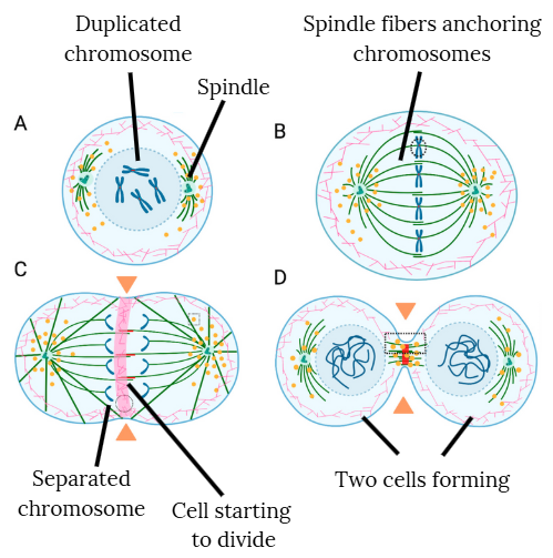
We can see this in the diagram below, where a single cell in step A divides into two by step D during meiosis II.
I’ll explain the steps in more detail (while intentionally keeping it simple!):
- In step A, the spindle starts to form. Note that the chromosomes look X-shaped because they’re duplicated and stuck together.
- These duplicated chromosome pairs are anchored in step B, then separated by the spindle fibers as they pull each single chromosome to opposite ends of the cell in step C.
- You can then see two cells form in step D, with the chromosomes becoming more squiggly looking as they loosen up and mix together.
At the heart of it, that’s how meiosis works. What you saw above is actually meiosis II, but the same basic steps, or stages, occur during meiosis I. These stages are known as prophase, metaphase, anaphase and telophase:
- Step A refers to the prophase stage, where the spindle forms.
- Step B refers to metaphase, where the chromosomes line up.
- Step C refers to anaphase, where the single chromosomes (aka chromatids) separate.
- Step D refers to telophase where the cells start to split.
And as I mentioned, there are two rounds of meiosis.
Meiosis II was shown above, but the same stages occur during meiosis I. Because there’s two rounds of meiosis, there’s also two rounds of prophase, metaphase, etc. and we refer to the prophase stage of meiosis I as prophase I, and prophase stage of meiosis II as prophase II!
I hate to throw all this lingo at you, but these specific stages are important, as we’ll see!
Meiosis I and II together!
Now let’s put it all together and look at both meiosis I and II together.
Before meiosis even begins, a stage called interphase takes place where the DNA is copied or replicated. This is how we get duplicated chromosomes at the beginning of meiosis I, which is why the chromosomes have an X-like shape. These are two identical chromosomes (called sister chromatids) that are stuck together, side by side, to give this shape.
This isn’t the same as the chromosome pairs that we’ve been talking about. The two copies of chromosome 1, for example, are slightly different and one copy comes from the egg and the other comes from the sperm. These are also known as homologous chromosomes and they both are duplicated before meiosis starts as shown below:

After the 23 pairs of chromosomes duplicate, meiosis is ready to start!
Remember, to make egg and sperm cells, the 46 chromosomes in our body cells are reduced to 23 by meiosis. At the beginning of meiosis I, there’s a single cell that has 46 chromosomes, and at the end of meiosis II, there are 4 cells with 23 chromosomes that are egg or sperm cells.
You can see both rounds and all the stages in the diagram below, which we’ll discuss in more detail.

The goal of meiosis I is to split up the chromosome pairs, so from a single cell with 23 pairs you get two cells that each have one copy of the 23 chromosomes.
- You begin with a single cell that has 46 chromosomes (23 pairs).
- Each chromosome pairs up with its matching partner (aka its homologous chromosome) — so the two copies of chromosome 1 pair up with each other. These pairs might exchange small bits of genetic material (this is called crossing over), which helps create genetic diversity (this is why the blue and red are mixed in some chromosomes). This structure of a pair of chromosomes stuck together is called a bivalent, and they’re stuck together by proteins called cohesins. We’ll talk about this more later.
- In metaphase I, the spindle anchors on to structures called kinetochores of the chromosome pairs. The pairs are then pulled apart in anaphase I, so each new cell will have 23 chromosomes (one from each pair).
- You end up with two cells, each with 23 chromosomes, but these chromosomes are still duplicated (with the X-like shape).
The goal of meiosis II is to separate the duplicated chromosomes, so from two cells with 23 duplicated chromosomes, you get 4 cells that have 23 unduplicated chromosomes.
- You begin with the two cells from meiosis I, each with 23 duplicated chromosomes.
- The X-shaped chromosomes are pulled apart into single strands (called chromatids).
- You end up with four cells, each containing 23 single chromosomes. These cells are either the sperm or egg cells.
And that’s meiosis! I hope that made sense. You can check out this video from Nucleus Biology on Youtube to put it together. If it doesn’t embed, here’s the link.
How meiosis lines up with egg development
Remember that you’re born with all the eggs you’ll have. These eggs are contained in very basic follicles called “primordial follicles” and represent your ovarian reserve. These eggs are locked in prophase I of meiosis and remain this way until puberty, at which point hormones allow one of these eggs to continue meiosis during ovulation. The egg then resumes meiosis I and continues its development by becoming an M1 (metaphase I) and then an M2 (metaphase II). The egg is locked into the metaphase II stage as an M2 egg, or mature egg, until it’s fertilized by a sperm.
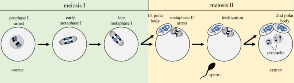
A diagram showing some of the steps of meiosis I and meiosis II. The blue squiggly lines inside the egg are chromosomes that are reduced in number as the egg develops. Modified from Clark and Akera (2021), CC BY 4.0
You’ll notice that there’s a polar body during the metaphase II stage. After meiosis I is finished, half of the chromosomes are separated to reduce the chromosome number, and these are placed in a polar body. The polar body is essentially a trash bin to dump excess chromosomes.
A second polar body is released at the end of meiosis II, which happens after the oocyte is inseminated and meiosis II resumes.
As we’ll see, problems with the spindle machinery can develop during the lengthy prophase I stage in the egg, so when meiosis resumes there can be errors in separating chromosomes.
Female age is strongly linked to embryo aneuploidy
With increasing female age, birth rates drop and miscarriage rates rise:

Aneuploidy has been reported in 50-70% of miscarriage products of conception and is considered the leading cause of miscarriages (Hyde et al. 2015). Maternal age is strongly linked to aneuploidy, and the chance of getting an aneuploid embryo increases with age:
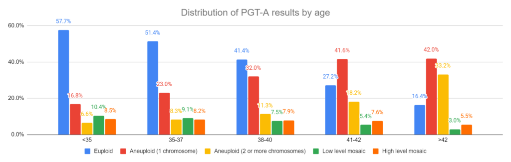
If you want to read more about the distribution of PGT results among IVF patients, check my post A look at how PGT-A results change with age, using data from over 86,000 biopsies.
So what about sperm?
Embryo aneuploidy can definitely occur due to sperm, but it’s about 10x more likely to be due to the egg. Bonus et al. (2022) used single nucleotide polymorphisms (SNPs) to identify parental origin of aneuploidies in embryos that were biopsied. They found that the majority of aneuploidies were of maternal origin:

Why is aneuploidy more commonly attributed to the egg, and why is this linked to age?
As mentioned in the previous section, most of the eggs are locked up in prophase I of meiosis and remain this way until puberty, at which point hormones allow one of the eggs to resume meiosis during ovulation. Sperm is “made fresh” continuously and doesn’t experience a pause in meiosis like this.
In older women, some of these eggs could have been paused in prophase I for decades!
It’s thought that one of the major causes of aneuploidy in eggs, especially in older women, is due to this extended period of prophase I. As we’ll see, different structures that are critical to meiosis can degrade with time, which explains why aneuploidy in the egg increases with age.
What causes aneuploidy?
Now let’s get to it and talk about how aneuploidy can actually occur!
There are several major ways that aneuploidy occurs, all of which are related to meiosis:
- Defects in the spindle
- Defects in cohesins
- Problems with maternal transcripts
- Genetic factors
- Mitochondrial dysfunction
Let’s look at these all in more detail!
Defects in the spindle
The spindle is responsible for pulling chromosomes apart during meiosis. Defects in how the spindle forms, or how it attaches, can lead to errors in separating the chromosomes. This can lead to some sperm or egg cells having the incorrect number of chromosomes.
The meiotic spindle anchors onto chromosome to separate them during meiosis. You can see this below. The spindle fibers are made of up of tube-like structures called microtubules (in green), and these physically attach to structures on the chromosomes (in blue) called kinetochores (in orange).
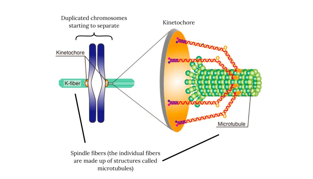
Once these spindles attach to the kinetochore, signals can be sent to tell the cell that that particular chromosome is properly anchored and is ready to be pulled apart and separated. Each of the 23 chromosomes have a kinetochore, and the spindle assembly checkpoint ensures these attachments are correct before allowing meiosis to proceed.
You can see how the spindle fibers (microtubules) attach to the kinetochore and begin chromosome separation in the video below (link, courtesy of WEHImovies on Youtube). This video shows chromosome separation during mitosis, and not meiosis, but the separation uses similar mechanics.
So how can this system be compromised in older women?
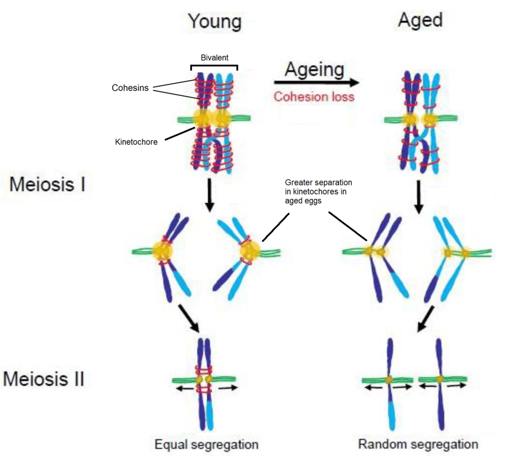
- As women age, the kinetochores can separate more (more on this in the next section), leading to errors in chromosome attachment during meiosis. This can cause chromosomes to attach to both spindle poles or misalign, increasing the risk of improper separation during cell division (Zielinska et al. 2015).
- In aged eggs, kinetochores and the parts of the chromosome they attach to can degrade. This leads to unstable, fragmented kinetochores that can attach incorrectly during meiosis, increasing the risk of abnormal chromosome numbers (Zielinska et al. 2019).
- In eggs, the spindle assembly checkpoint is less strict compared to other cells, making them more prone to chromosome segregation errors during meiosis (Wartosch et al. 2021). Additionally, this checkpoint is compromised in older women (Cimadomo et al. 2018) which can result in higher errors in separating chromosomes.
Defects in cohesin
As mentioned, eggs are paused in prophase I until ovulation. During this period, which can last decades, pairs of chromosomes are held together in a bivalent structure.

This bivalent structure is maintained for this long period by ring-like protein complexes called cohesins, which wrap around chromosomes together until they are ready to separate when meiosis resumes during ovulation. As women age, the levels of cohesins decrease, which is thought to be a major factor in the rise of aneuploidy with age (Wartosch et al. 2021).
As cohesins are lost, the bivalent structure isn’t held together as strongly. This can lead to the kinetochores separating more, which can serve as an additional site for the spindle to attach to, potentially causing aneuploidy.
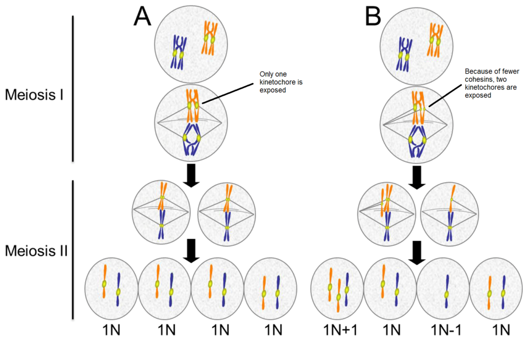
You can see how this can lead to aneuploidy below. Normal meiosis in younger eggs is seen in (A) and incorrect meiosis with older eggs in (B). In A, only a single kinetochore is exposed because of how tightly the cohesins are holding the bivalent together. In B, the weaker cohesins allow the kinetochores on one duplicated chromosome to separate more, so the spindle can erroneously attach to both of them. This can lead to the wrong number of chromosomes in the eggs (in this example, 2 of 4 eggs are aneuploid).
This research is mainly based on mice and it’s still uncertain whether human eggs lose cohesin in the same way (Wartosch et al. 2021). However, it’s been reported that the structure of bivalents can change as women age, especially with smaller chromosomes like 21 and 22. Bivalents for these chromosomes are more likely to become unpaired because they’re smaller and more susceptible to reduced cohesins. This might explain why these chromosomes are commonly aneuploid in older women.
Maternal transcripts
Before an embryo’s genome activates around day 3 of development, it relies on maternal factors stored in the egg to manage its early growth. These factors, including RNA transcripts and proteins, are like a pre-packed toolkit, ensuring the embryo has everything it needs before it can produce its own on day 3. You can read more about embryonic genome activation in my post Embryo arrest.
One key aspect of these maternal factors is their role in meiosis. As women age, the regulation of these stored transcripts and proteins might be compromised (Zielinska et al. 2019). For instance, proteins essential for accurate chromosome segregation may not be produced correctly from these transcripts, increasing the risk of errors like aneuploidy.
Genetics and aneuploidy
While the risk of aneuploidy increases with maternal age, genetic factors also play a role in determining aneuploidy rates in IVF patients. Research has shown that some women, regardless of age, can be genetically predisposed to producing embryos with aneuploidy.
Specific genes have been implicated in influencing aneuploidy:
- Mutations in genes TUBB8, PATL2, and WEE2, can predispose younger women to higher rates of aneuploidy (Wartosch et al. 2021). TUBB8 encodes a protein involved in forming microtubules of the spindle; PATL2’s function isn’t clear, but may be involved in processing maternal transcripts; WEE2 is involved in keeping the egg paused in prophase I.
- Genome analysis of parents who had children with trisomy 21 (Down syndrome) showed that some genes may be involved in this type of aneuploidy (Chernus et al. 2019). Several of the genes were involved in meiosis, including AURKC, a gene that codes for Aurora kinase C that’s involved in the spindle assembly checkpoint.
- Nguyen et al. (2017) sequenced the AURKB and AURKC genes in patients with higher or lower rates of aneuploid embryos. They found some patients had one variant of AURKC that predisposed them to higher rates of aneuploidy, but they also identified a protective variant of AURKB in one particular woman, who had lower than average aneuploidy rates! This protective variant allowed the Aurora kinase B protein to better regulate the alignment of chromosomes during metaphase, so it actually decreased the chance of aneuploidy.
- Whole-exome sequencing of women with more than 50% aneuploid blastocysts revealed a number of gene variants that predisposed them to aneuploidy (Tyc et al. 2020). One of the genes, CEP120, is involved in microtubule formation of the spindle.
- Wu et al. (2024) found that patients with mutations in HAUS6, KIF11 and KIF18A had increased risk of aneuploidy, along with embryo arrest, egg maturation problems and fertilization failure. These genes code for proteins involved in spindle assembly.
Mitochondria may be involved in aneuploidy, too
During meiosis, many different cellular components participate to separate the chromosomes. Obviously, this process requires energy!
In the egg, and other cells, energy comes from cellular organs called mitochondria. These are like the batteries of the cell and supply energy when needed. They have their own DNA, called mitochondrial DNA, which provides instructions for mitochondrial components.
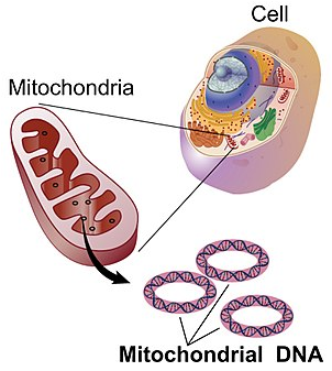


Over time, mitochondria accumulate mutations in their DNA and sometimes those mutations occur in critical genes that are needed for proper mitochondrial function. This can result in the mitochondria not producing enough energy for the cell (Cimadomo et al. 2018), which can cause problems with spindle assembly during meiosis (Zhang et al. 2023). This could lead to increased risk of aneuploidy.
Mitochondria are also known to be a major producer of reactive oxygen species (ROS), leading to oxidative stress in the cell. This stress can damage the spindle apparatus and disrupt chromosome alignment, increasing the risk of aneuploidy. However, antioxidants like melatonin have been shown to lower this risk by neutralizing ROS and improving mitochondrial function in mice, leading to proper spindle formation and chromosome segregation (Zhang et al. 2022).
What are the most common aneuploidies?
Wartosch et al. (2021) compiled data on which chromosomes were the most commonly affected in a variety of situations. These all involved trisomies, or three copies of a chromosome (ie. +1 means trisomy 1). Having an extra copy of a chromosome can disrupt the normal “dosage” of gene expression, which can lead to developmental problems. The reason we don’t see any monosomies here, where there’s only one copy of a chromosome, is because these typically aren’t compatible with life.
- For eggs, the most common aneuploidies in young women are +1, +2, +3, +4 and +5.
- For eggs, the most common aneuploidies in older women are +13, +14, +15, +16, +21 and +22.
- For sperm, the most common aneuploidies are +13, +15, +21, +22, and those involving the sex chromosomes (X and Y).
- For cleavage stage embryos, the most common aneuploidies are +15, +16, +21, +22.
- For blastocysts, the most common aneuploidies are +15, +16, +21, +22.
- In pregnancy loss, the most common aneuploidies are +13, +15, +16, +18, +21, +22.
- For stillbirths, the most common aneuploidies are +13, +18, +21, and those involving the sex chromosomes (X and Y).
- For live births, the most common aneuploidies are +13, +18, +21, and those involving the sex chromosomes (X and Y).
A 2024 study investigated the genetics of over 3,200 miscarriages, finding that two-thirds were associated with a chromosomal abnormality, with +16 and the 8p23.1 deletion as the most common abnormalities. Other common aneuploidies included +22, +21 and +15, with +22 and +15 being particularly common in women >35. You can read more about this in my post Study performs genetic analysis on over 3,200 miscarriages.
Can I transfer an aneuploid embryo?
Often patients who do PGT-A wonder about whether they can transfer an aneuploid embryo.
Generally, aneuploid embryos are not recommended for transfer.
There isn’t much data on it, but one study transferred untested embryos that were biopsied, and the biopsy results weren’t revealed until after the pregnancy results. Of the 102 aneuploids that ended up being transferred, none of them led to a live birth, and all either failed to implant or miscarried. You can check out the full summary in my post Transferring aneuploid embryos.
In another study, 76 non-complex aneuploids (with <3 affected chromosomes) were transferred, resulting in one live birth. The aneuploid embryo that led to a live birth was originally tested as 46 XX +14, -18 but a euploid female was delivered. You can read more about this in my post 144 “abnormal” (aneuploid/mosaic) embryos and their outcomes.
So why do aneuploid embryos sometimes lead to healthy live births?
It’s possible that the aneuploid embryo isn’t truly aneuploid — it could be a mosaic!
A PGT-A biopsy takes only 5-10 cells from an embryo, which can be hundreds of cells large, and this small sample may not be truly representative of the rest of the embryo. Furthermore, this biopsy is taken from the trophectoderm which develops into the placenta, and not the inner cell mass (ICM) which develops into the fetus.
So it’s possible that some “aneuploids” are just mosaics, and they contain some euploid cells as well. For example, if 90% of the cells of an embryo are aneuploid, and only aneuploid cells are taken for the biopsy, then the result would be aneuploid. A repeat biopsy might take some of the euploid cells, which would give a different result.
Mosaic embryos can “self-correct,” where the euploid cells outgrow the aneuploid cells (read more about that in my post on mosaics). This process can give rise to a euploid fetus from a mosaic embryo. Embryos that are fully aneuploid likely don’t self-correct like mosaics.
How often do aneuploids retest as aneuploid?
In one study, researchers retested 1,117 aneuploids, finding that 81.4% were concordant and tested aneuploid a second time. Another study retested aneuploids up to 4 times (including the ICM), finding that the results were the same 98% of the time, while another study found that retesting confirmed aneuploidy 97% of the time.
So in general, aneuploid results indicate that there’s a high chance the embryo is fully aneuploid, although there’s still a chance that you can get a different result. Retesting mosaics tended to give variable results. You can read more about this in my post Does a PGT-A biopsy match the rest of the embryo?
Conclusions
Aneuploid embryos, which have an abnormal number of chromosomes, are a major cause of miscarriages and implantation failures.
Aneuploidy occurs when chromosomes separate incorrectly during meiosis, the process that reduces chromosome numbers from 46 to 23 in egg and sperm cells. This separation is controlled by the spindle apparatus, a cellular structure that anchors to chromosomes and pulls them apart.
Errors in spindle attachment can lead to improper chromosome separation, resulting in aneuploidy, where sperm or egg cells have the wrong number of chromosomes. Aneuploidy is closely linked to female age because eggs are paused in meiosis for decades, during which structures like cohesins and kinetochores deteriorate, increasing the risk of spindle errors.
Some women are more likely to produce aneuploid embryos due to specific genetic mutations that compromise key structures needed for successful meiosis. Interestingly, some mutations can be beneficial; for example, one woman was found to have a mutation that actually reduced her risk of embryo aneuploidy.
Generally, aneuploid embryos are not recommended for transfer due to their low likelihood of resulting in a live birth, although some may actually be mosaic and could lead to a healthy pregnancy.
About Embryoman
Embryoman (Sean Lauber) is a former embryologist and the founder of Remembryo, an IVF research and fertility education website. After working in an IVF lab in the US, he returned to Canada and now focuses on making fertility research more accessible. He holds a Master’s in Immunology and launched Remembryo in 2018 to help patients and professionals make sense of IVF research. Sean shares weekly study updates on Facebook, Instagram, and Reddit regularly. He also answers questions on Reddit or in his private Facebook group.