
A 2024 study investigated the genetics of over 3,200 miscarriages, finding that two-thirds were associated with a chromosomal abnormality, with trisomy 16 and the 8p23.1 deletion as the most common abnormalities.
Miscarriage can be caused by a number of factors, with chromosomal abnormalities accounting for the majority.
From over, 3,200 miscarriages, this study measured both numerical chromosomal abnormalities (changes in the number of chromosomes, like trisomies and monosomies) and structural chromosomal abnormalities (changes in segments of a specific chromosome) in the miscarriage. They also looked at how these abnormalities changed with age, repeat miscarriage and whether the miscarriage occurred before or after 12 weeks.
To better understand the results of this study, you should check out how to understand PGT-A reports. While the study presented here doesn’t involve euploid transfers or PGT-A, it does discuss chromosomal abnormalities.
🔗 Original studies are referenced in this post or within the linked Remembryo posts.
💡 Reminder: Terms underlined with a dotted black line are linked to glossary entries. Clicking these does not count toward your paywall limit.
Study details
This section covers key details of how the study was performed, including patient characteristics, how they were treated, and other methods used. For those who aren’t interested in these details, and just want to see the results, you can go ahead and skip this part.
- This was a retrospective study that took place at a single hospital in China between 2015 and 2023.
- All included women had a miscarriage before 28 weeks, and the miscarriage tissue (products of conception) were genetically tested.
- Genetic testing used copy number variation sequencing (CNV-seq) using NGS technology, while short tandem repeat analysis was done to detect polyploidy, uniparental diploidy (UPD) and maternal cell contamination.
- Chromosomal abnormalities were identified using the 2019 guidelines from the American College of Medical Genetics.
In terms of sample size, there were a total of 3,247 miscarriages with products of conception that were genetically tested (2,756 cases of chorionic villus and 491 cases of fetal tissue), which included the following subgroups:
- Age: <35 (2,551 women), ≥35 (696 women)
- History of miscarriage: one miscarriage (1,690), more than one miscarriage (1,557).
- Gestational age: <12 weeks (2,871), ≥12 weeks (376)
Miscarriages with a numerical chromosomal abnormality
Of the 3,247 miscarriages with products of conception that were genetically tested, 2,169 had chromosomal abnormalities (66.8%):
- 45.2% (1,467) had numerical chromosomal abnormalities (abnormalities in the number of chromosomes).
- 21.6% (702) had structural chromosomal abnormalities (abnormalities in the structure of chromosomes).
In this section we’ll focus on the numerical chromosomal abnormalities.
Of the 1,467 miscarriages with a numerical chromosomal abnormality:
- 40.1% (1,302) involved aneuploidy.
- 5.1% (165) involved polyploidy (more than 2 complete sets of chromosomes; more than 97% were triploid).
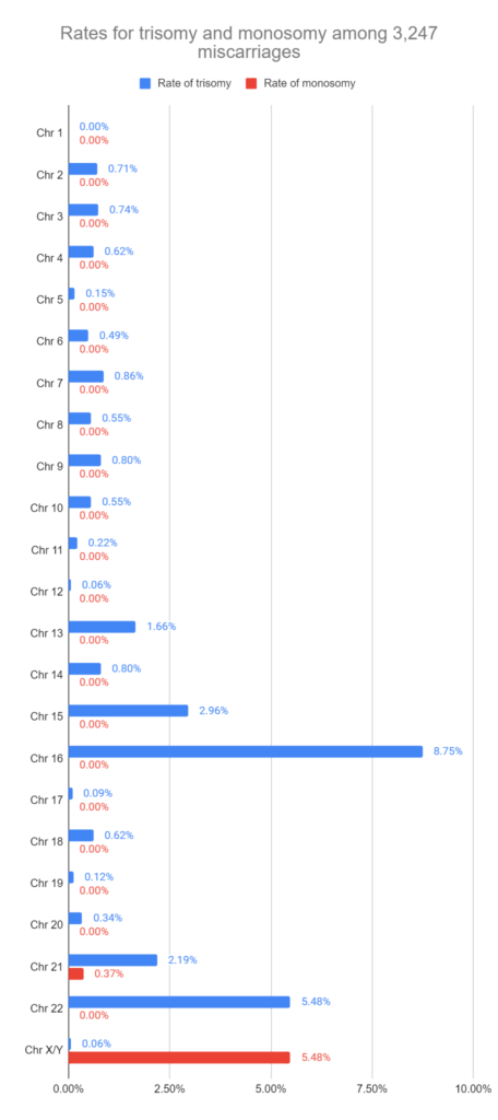
For the 1,302 miscarriages that had aneuploidy, 1,126 had a single aneuploidy. This means that the miscarriage tissue had only a single chromosome affected. The vast majority of single aneuploid miscarriages were trisomies, meaning there was an extra copy of a chromosome, while there were very few monosomies (presumably these don’t survive past the embryo stage). The most common trisomy in the miscarriages was trisomy 16 (8.75%), followed by trisomy 22 (5.48%) and trisomy 15 (2.96%). You can see the distribution below:
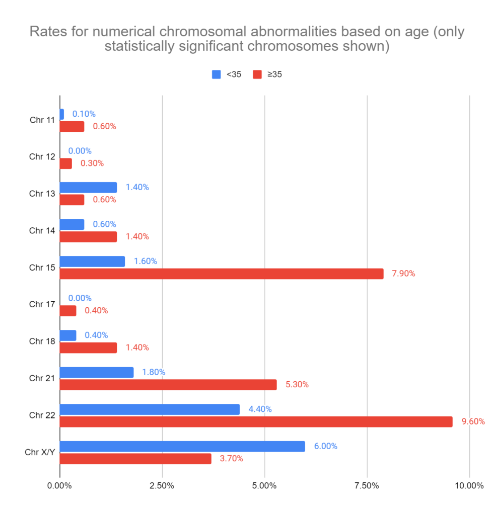
They also grouped the miscarriages based on age (<35 vs ≥35), miscarriage history (1 vs more than 1) and gestational age (<12 weeks vs ≥12 weeks).
For age, there were quite a few differences. Overall, there were more numerical chromosomal abnormalities in miscarriages from women ≥35 (41.3% vs 59.5%, p< 0.001). Only the statistically significant differences for specific chromosomes are shown in the graph below, while the rest of the chromosomes had a similar rate of numerical abnormalities (and similar to what was seen above).
For miscarriage history (1 vs more than 1), only chromosome 6 showed a statistical difference (0.2% vs 0.8%).
For gestational age (<12 weeks vs ≥12 weeks), there were more numerical chromosomal abnormalities in early miscarriages that happened before 12 weeks (46.7% vs 33.8%, p< 0.001). For specific chromosomes, the following showed significant differences:
- Chromosome 16 (9.4% vs 3.5%)
- Chromosome 18 (0.5% vs 1.6%)
- Chromosome 21 (2.3% vs 4.2%)
- Chromosome 22 (5.8% vs 2.9%)
- Chromosome X/Y (5% vs 9.8%)
They also looked at rates of mosaicism between these 3 groups, without any differences (all occurred at a frequency between 3-4%). The overall rate of mosaicism based on all the miscarriages was 4.7%.
The products of conception that were genetically tested in this study were taken from the chorionic villus and fetal tissue. There was a higher degree of numerical aneuploidy in chorionic villus samples compared to fetal tissue (48.7% vs 25.7%, p< 0.001). There was also a higher degree of mosaicism (4% vs 1.8%).
Miscarriages with a structural chromosomal abnormality
Of the 3,247 miscarriages with products of conception that were genetically tested, 2,169 had chromosomal abnormalities (66.8%):
- 45.2% (1,467) had numerical chromosomal abnormalities (abnormalities in the number of chromosomes).
- 21.6% (702) had structural chromosomal abnormalities (abnormalities in the structure of chromosomes).
In this section we’ll focus on the structural chromosomal abnormalities.
Unlike numerical chromosomal abnormalities that affect the number of whole chromosomes, structural chromosomal abnormalities refer to abnormalities in parts of chromosomes, including deletions, duplications, inversions and translocations.
In this study they categorized the structural chromosomal abnormalities as pathogenic or likely pathogenic, which means they have a strong association with an increased risk of miscarriage.
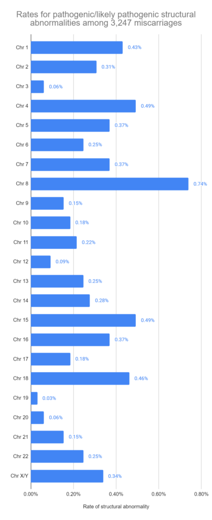
Of the 702 miscarriages that had structural chromosomal abnormalities, 212 were pathogenic or likely pathogenic. The rest were mostly of “uncertain significance,” with a small number being benign or likely benign. You can see the distribution of the 212 pathogenic/likely pathogenic structural abnormalities below, which includes both duplications and deletions.
They also grouped the miscarriages based on age (<35 vs ≥35), miscarriage history (1 vs more than 1) and gestational age (<12 weeks vs ≥12 weeks). There was a higher degree of structural abnormalities in younger women (23.2% vs 15.9%, p<0.001), but no differences in miscarriage history or gestational age.
You can see the most affected regions for the pathogenic or likely pathogenic structural abnormalities below, along with their frequency (over all the miscarriages in this study) and some information on what they’re linked to.
- 8p23.1 deletion: 0.40% – linked to stillbirth, early miscarriage, cardiac abnormalities.
- 18p11.32p11.31 deletion: 0.22% – linked to Chromosome 18p deletion syndrome.
- 4p16.3p16.1 deletion: 0.22% – linked to Wolf-Hirschhorn syndrome.
- 5p15.33p15.2 deletion: 0.22% – linked to cri du chat syndrome.
- 8q24.22q24.3 duplication: 0.22% – linked to missed miscarriage.
- 1p36.33p36.32 deletion: 0.15% – linked to miscarriage.
- 11q24.3q25 duplication: 0.15% – linked to miscarriage.
- 15q23q25.2 duplication: 0.15% – linked to miscarriage.
- 18q21.33q22.2 deletion: 0.15%
- 15q11.2 deletion: 0.15%
- 16p13.11 duplication: 0.15%
- 7p22.3p22.2 deletion: 0.12%
- 4p16.3p15.31 duplication: 0.12%
- 6q25.3q27 duplication: 0.09%
- 15q26.2q26.3 deletion: 0.09%
- 15q26.1 deletion: 0.09%
- 4q32.3q35.2 deletion: 0.09%
- 7q36.3 deletion: 0.09%
- Xp22.31 deletion: 0.09%
- 1p36.31p36.12 duplication: 0.09%
- 2q36.3q37.3 duplication: 0.09%
- 6q14.1 duplication: 0.09%
They compared the frequency of these regions to other cohorts (from outside this study), including the general population and an early miscarriage cohort. Nearly all the regions above were found in the early miscarriage cohort.
Further analysis and refinement of the above regions led to the identification of 86 genes that were associated with miscarriage: 3 of these genes (PARP6, ISLR, ULK3) are involved in early embryo development, while 4 genes (FGFRL1, TBC1D14, SCRIB, PLEC) are involved in early miscarriage.
Conclusions
This study found that the overall rate of chromosomal abnormalities in miscarriage was 66.8%, while the overall rate of mosaicism was 4.7%.
For numerical chromosomal abnormalities, the vast majority were trisomies, with only monosomies found for chr 21 and X/Y. Trisomy 16 was the most common numerical chromosomal abnormality found in miscarriages.
For structural chromosomal abnormalities, chr 8 was most affected in miscarriages, with the 8p23.1 deletion being the most common. The authors identified 86 genes involved in miscarriage, such as PARP6 and ULK3 that are involved in early embryo development.
They also grouped the miscarriages based on age (<35 vs ≥35), miscarriage history (1 vs more than 1) and gestational age (<12 weeks vs ≥12 weeks).
For age, there were more numerical chromosomal abnormalities in older women, and more structural abnormalities in younger women. Chr 22 was the most affected chromosome in miscarriages from older women.
For miscarriage history, numerical abnormalities in chr 6 were slightly more common with repeat miscarriages, with no differences in structural abnormalities.
For gestational age, there were more numerical abnormalities in chr 16 and 22 for early miscarriage, and more numerical abnormalities in chr 18, 21 and X/Y for late miscarriages. There were no differences for structural abnormalities.
Related studies
To learn more about this topic, you can check out a number of studies referenced in this study below (5 links):

Reference

About Embryoman
Embryoman (Sean Lauber) is a former embryologist and the founder of Remembryo, an IVF research and fertility education website. After working in an IVF lab in the US, he returned to Canada and now focuses on making fertility research more accessible. He holds a Master’s in Immunology and launched Remembryo in 2018 to help patients and professionals make sense of IVF research. Sean shares weekly study updates on Facebook, Instagram, and Reddit regularly. He also answers questions on Reddit or in his private Facebook group.
Related posts:
 Meta-analysis combines 74 studies to examine factors linked to euploid transfer success
 Study compares PGT-A outcomes for patients with different diagnoses
 ASRM’s 2024 committee opinion on the use of PGT-A
 Small genetic mutations in euploid pregnancies linked to miscarriage
 PGT-A miscarriage rates (euploid, mosaic, and aneuploid transfers)
 Pilot study shows improved IVF outcomes after spindle transfer in women with poor egg quality
 Euploid embryos show reduced implantation potential with advancing maternal age
 Can machine learning identify a euploid miscarriage without PGT-A?