A 2024 study performed a more sensitive type of PGT-A by analyzing single cells, finding that nearly all blastocysts and fetal tissue contain some level of mosaicism.
PGT-A is commonly used in IVF to select embryos with the best chance of success. One of the problems with PGT-A is that it uses a single biopsy of about 5-10 cells to represent the whole embryo, which could be hundreds of cells in size.
Embryos with both euploid and aneuploid cells are called mosaic. However, determining the true incidence of mosaicism is difficult, because the small number of cells biopsied may not be representative of the whole embryo.
Another problem with PGT-A is that the whole 5-10 cell biopsy is DNA sequenced together as one sample. This could mask the results of individual cells and make detecting mosaicism more difficult.
This post is a summary of a study by Zhai et al. (2024), who used a more sensitive form of PGT-A to sequence individual cells from embryos/fetuses to see how common mosaicism is. They sequenced cells from 3 different stages:
- Pre-implantation: From blastocysts donated by couples who had IVF.
- Post-implantation: From embryos that were cultured in an experimental model that mimicked implantation to day 8-14.
- Fetus: From cells from miscarriage tissue, including the cerebral cortex, heart and kidneys. All of these cells were from embryos/fetuses between 5-26 weeks gestation.
It’s important to point out that we can’t use single cell sequencing to do PGT-A — it requires that DNA is extracted from each cell in the embryo, which kills all the cells in the process. The point of this study is to see how common mosaicism really is.
For more background on PGT-A, check out my Complete guide to PGT-A.
🔗 Original studies are referenced in this post or within the linked Remembryo posts.
💡 Reminder: Terms underlined with a dotted black line are linked to glossary entries. Clicking these does not count toward your paywall limit.
All pre-implantation blastocysts showed mosaicism
In this study, 20 blastocysts were separated into 1133 single cells, which were then individually DNA sequenced.

You can see the details for each blastocyst below, including the age of the patient, the embryo grade and how many cells were harvested. Not all the cells could be sequenced, so there’s a smaller number in the “analyzed” row.
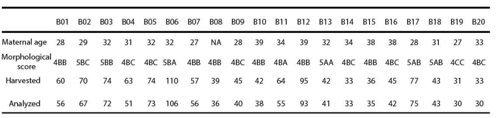
Of the 20 blastocysts that had single cell sequencing, all 20 had some level of mosaicism. On average, the blastocysts had 25% mosaicism per embryo. You could see the % mosaicism for each blastocyst below (the red bar), ranging from about 15% for sample B16 to about 95% for B01.

In about 70% of the blastocysts, there was “complementary aneuploidy” as illustrated below. Complementary aneuploidy refers to a condition where some cells have an extra chromosome while others have one less. This can happen due to errors in mitosis, the type of cell division that embryos use to grow and make more cells. During mitosis, a cell doubles its DNA and chromosomes and then splits into two new cells.
In the example shown below, one cell of a 2 cell embryo might make an error where chromosome 4 isn’t separated properly during mitosis. This results in one cell with an extra copy of chromosome 4 (+4) and one cell with a missing copy of chromosome 4 (-4). These cells then divide further as the embryo grows, so the blastocyst has a mix of +4 and -4 cells.

So why does this matter? Traditional PGT-A involves the DNA sequencing of a single 5-10 cell biopsy together as a single sample, and this can underestimate mosaicism in embryos with complementary aneuploidy. Let’s look at an example:
- Let’s say we DNA sequence a whole 10 cell biopsy of the blastocyst in the example above, and 6 cells are normal, 2 have an extra chromosome 4 (+4), and 2 are missing chromosome 4 (-4). With standard PGT-A, the whole 10 cell biopsy is DNA sequenced together, and the overall result would appear normal because the extra and missing chromosomes balance each other out.
- However, with single-cell PGT-A, each cell is analyzed individually, revealing that 20% of cells have an extra chromosome and 20% are missing one, highlighting the presence of mosaicism. This suggests that mosaicism in many blastocysts may be underestimated using standard PGT-A methods, especially since 70% of blastocysts exhibit complementary aneuploidy.
Nearly all post-implantation blastocysts showed mosaicism
Next, the researchers examined embryos that developed from day 8-14 in an experimental model that mimicked implantation. This involved placing blastocysts onto an specialized substrate that resembled the uterine environment. The embryos attached and grew more advanced structures until day 14, at which point the experiments were terminated for ethical reasons.
This analysis involved 4820 cells from 28 embryos at the day 8-14 stage. Mosaicism was detected in 27 out of 28 of the embryos (96.43%), which was mostly present in the trophectoderm cells.
The fact that they found more trophectoderm cells carrying these chromosomal errors suggests a self-correction mechanism, which was also shown in another study using a mosaic embryo model. In this study, the researchers showed that aneuploid cells were more likely to differentiate (change) into trophectoderm cells, allowing the ICM to become enriched in euploid cells. You can read more about this in my post Depletion of aneuploid cells in mosaic embryos.
Nearly all fetal tissue showed mosaicism
Finally, the researchers analyzed single-cell PGT data from embryos/fetuses at 5-26 weeks gestation, which was from the author’s previous studies. Single cells from cerebral cortex (brain), heart and kidney cells were isolated from miscarriage tissue and DNA sequenced (48 samples in total: 19, 18 and 11 respectively).
The researchers observed that 47 out of 48 samples exhibited mosaicism (97.92%). Specifically, the cerebral cortex samples displayed an average mosaicism rate of 4.70%, while the hearts showed 12.46% and the kidneys demonstrated 11.98%. This suggests that the heart and kidney tissue can tolerate higher levels of mosaicism than the brain. You can see the results for the tissue samples below (red bar indicates % mosaic).

A second PGT biopsy shows that even some euploid embryos are mosaic
One last thing!
The researchers at their IVF clinic had a habit of splitting their PGT-A biopsy into two: One sample was submitted for PGT-A, while a second “backup” sample was frozen and used if needed.
For the embryos that were euploid and led to live births, they sequenced the backup biopsy sample to see if it matched the first one.
Of the 116 samples, 8 of the backups were completely aneuploid and 4 were mosaic. So 12 of 116 (10.34%) of the “euploids” were actually mosaic. Remember, the first euploid result counts too, so even if the result is mosaic or aneuploid in the backup sample, the overall result is mosaic.
In truth, most (all?) of these euploids were probably mosaic, but in this experiment they were limited to only testing the 5-10 cells of a backup biopsy.
Regardless, all 116 euploids resulted in healthy live births, even in the 8 embryos that would have been considered aneuploid if only the backup biopsy was tested. This just shows that sampling a single biopsy for PGT-A can be flawed, as it may not represent the whole embryo.
This isn’t new information, I’ve reviewed a number of studies that have shown that retesting euploid or aneuploid embryos can reveal that the embryo is actually mosaic (check out my post Does a PGT-A biopsy match the rest of the embryo?).
Conclusions
This study used a more sensitive form of PGT-A to sequence up to 100 individual cells from embryos and fetuses to see how common mosaicism is.
They found that nearly all of these samples showed some level of mosaicism: 100% of blastocysts, 96% of day 8-14 post-implantation embryos and 98% of fetal tissues (derived from 5-26 week miscarriages).
They also found that most blastocysts showed “complementary aneuploidy,” involving a gain and complementary loss of a chromosome in cells, potentially leading to reduced detection of mosaicism with standard PGT-A methods.
This isn’t the only study to do single cell sequencing of many blastocyst cells. Another 2024 study did something similar with 79 blastocysts, and found that 82% of them were mosaic to some degree. You can read more about that in my post Mosaicism much more common than previously thought, using more sensitive PGT-A technique.
While these results are really interesting, it’s important to note that this study involved single-cell sequencing on many blastocyst cells, which destroys the cells, so it can’t be used for clinical use (because it would kill the embryo). The point was to show how common mosaicism is in donated blastocysts and other tissues.
Reference

About Embryoman
Embryoman (Sean Lauber) is a former embryologist and the founder of Remembryo, an IVF research and fertility education website. After working in an IVF lab in the US, he returned to Canada and now focuses on making fertility research more accessible. He holds a Master’s in Immunology and launched Remembryo in 2018 to help patients and professionals make sense of IVF research. Sean shares weekly study updates on Facebook, Instagram, and Reddit regularly. He also answers questions on Reddit or in his private Facebook group.