Embryo arrest is when an embryo stops developing, usually before reaching the blastocyst stage. This post explains possible causes, including embryonic genome activation, maternal effect genes, the subcortical maternal complex, aneuploidy, mitochondrial dysfunction, oxidative stress, lab conditions, and sperm DNA fragmentation.
One post that I highly recommend you check out before reading this is my Biology 101 post that explains how genes are converted into proteins. This post doesn’t count toward the paywall’s limit.
🔗 Original studies are referenced in this post or within the linked Remembryo posts.
💡 Reminder: Terms underlined with a dotted black line are linked to glossary entries. Clicking these does not count toward your paywall limit.
Table of Contents
What is embryo arrest?
Embryo arrest is when an embryo stops developing for at least 24 h, typically during the cleavage stage or around day 3 (Sahin et al. 2023). This is the stage before the blastocyst stage, so arrested embryos don’t make it to blastocyst.
About 50-70% of embryos fail to make it to the blastocyst stage (Wong et al. 2010) and about 40% of all patients show at least one arrested embryo per cycle (Betts et al. 2008).
So “embryo arrest” is a fairly common phenomenon.
When an embryo arrests, it seems to enter a kind of senescent-like state where the cells stop dividing. A related term for this is “cellular senescence” and embryos that arrest are in a senescent-like state.
The only way to tell that an embryo has arrested is by comparing its development over time. Embryos grow pretty quickly and can double their cells in about 24 hours. If an embryo isn’t showing signs of cell division after 24 hours it’s a good sign that the embryo has arrested.

Some clinics will take embryos to day 6 before they discard arrested embryos. Embryos should become a blastocyst by day 5 or 6, so if an embryo is still at the day 3 cleavage stage by day 6 then some labs will assume it’s arrested.
Embryonic genome activation
An important cause for embryo arrest has to do with “embryonic genome activation.” This is when the genome (DNA) of the embryo becomes activated, usually around day 3 or so, and if this process fails then the embryo can arrest.

After fertilization, the sperm’s and egg’s DNA combine to form the embryo’s DNA, or its genome. Genome is the word we use to describe the complete set of DNA of an organism. It contains all the instructions for the embryo to produce the factors it needs to divide its cells and develop.
The embryo’s genome isn’t actually turned on until about day 3 when the embryo has about 8 cells (Hanna et al. 2018).
So if the genome isn’t activated until day 3, how does the embryo develop on days 1 and 2?
Before embryonic genome activation, the embryo relies on maternal factors that were stored in the egg before fertilization. This is kind of like when your parents would go away for the weekend and leave the fridge stocked with goodies.
Because the embryo’s DNA isn’t active yet, it can’t make its own factors, so it needs to rely on stored maternal factors.
These stored maternal factors are involved in a variety of processes and if the egg is missing these factors this can result in embryo arrest. For example, some factors may be involved in cell division, and if the egg is missing these then it won’t be able to divide during the early stages and will stop developing.
Let’s talk about these stored maternal factors in a bit more detail, starting with describing these factors more accurately as “RNA transcripts” and proteins. For those who aren’t familiar with these terms, I really encourage you to check out my Biology 101 post where I explain it all (it doesn’t count toward the paywall’s post limit).
Maternal effect genes
Stored egg factors include RNA transcripts and proteins. More broadly, the genes that encode these factors are called “maternal effect genes.”
Here’s some of the types of maternal RNAs and their function (with the corresponding gene in brackets) found in the early embryo (Wong et al. 2010):
- Involved in cell division (ANLN, AURKA, CFL1, ECT2, FGFR1)
- Energy production (ATP2C1)
- Transcription of DNA to make RNA (TAF4, TBP)
- Processing of RNA (CPEB1, PARN, SYMPK, DICER1)
- DNA reprogramming (EHMT2, DNMT3B)
Wong et al. (2010) found that transcripts involved in cell division and processing of RNA were all reduced in arrested embryos compared to normal embryos.
As an example, let’s say the embryo was missing ANLN (underlined above). This is the “anillin, actin binding protein” gene and is involved in cell division. Without it, the early embryo wouldn’t be able to divide and would arrest.
There have been 82 maternal effect genes identified in mammals as of 2022 (Mitchell 2022). Not all of them are necessarily involved in embryo arrest, but these are genes that have RNA transcripts/proteins that are stored in the egg.
Many of these have been identified in mice, however a few have been identified in humans. The genes that are in bold and underlined have been identified as being in humans by Mitchell 2022:
- Ago2
- Arida
- Atg5
- BCAR4c
- Bcas2
- Bnc1
- Btg4d
- Cdc20d
- Cdx2
- Cnot6l
- Ctcf
- Ctnnb1
- Cxxc1
- Dcaf13
- Ddb1
- Dgcr8
- Dicer1
- Dnmt1
- Dnmt3a
- Dnmt3l
- Dppa3
- Dtl
- Eed
- Ehmt2
- Exosc10
- Ezh2
- Gclm
- H3f3b
- Hira
- H2ac1
- H2bc1
- Hsf1
- Hsp90b1
- Igf2bp2
- Kdm1a
- Kdm1b
- Kdm4a
- Khdc3
- Kmt2d
- Kpna6
- Mapk1
- Mapk3
- Med12
- Mgat1
- Mlh3
- Nlrp2d
- Nlrp4f
- Nlrp5d
- NLRP7
- Nlrp9a,b,c
- Npm2
- Oeop
- Padi6d
- Patl2d
- Pdk1
- Plag1
- Pum1
- Rad9a
- Ring1
- Rnf2
- Setd2
- Setdb1
- Slbp
- Smarca4
- Tcl1
- Tet3
- Tle6d
- Trp73
- Trim28d
- Trip13
- TUBB8d,e
- Tut4
- Tut7
- Ube2
- Uchl1
- Uhrf1
- Yap1
- Ythdf2
- Zar1
- Zbed3
- Zfp36l2
- Zfp57
Large sequencing studies have identifyied genetic causes behind repeated embryo arrest or IVF cycles that produce no transferable embryos:
- Chen et al. (2025) examined women with unexplained infertility or repeated IVF failure and found that about 13% had mutations in genes involved in egg maturation, fertilization, or early embryo development, most commonly TUBB8, PATL2, PADI6, and NLRP5. Many of these genes control processes that occur before the embryo’s own genome becomes active, meaning defects can disrupt fertilization or cause embryos to arrest early. Read more in my post Scientists identify genes linked to unexplained infertility and repeated IVF failure.
- Zhang et al. (2025) focused on patients with repeated IVF cycles producing no transferable embryos and found detectable genetic causes in about 13–23% of cases, again involving genes linked to egg maturation and early embryo development, including TUBB8, TLE6, PADI6, and NLRP5. Read more in my post New genetic patterns found in women with low egg quality and no transferable embryos.
The subcortical maternal complex
The subcortical maternal complex (SCMC) is a multi-protein complex involved in embryonic genome activation.
Some of the protein members are KHDC3L, NLRP2/5/7, OOEP, PADI6, and TLE6. If you check the list of maternal effect genes above you’ll see some of these listed.
We’re not really sure of all the functions of the SCMC, but it is involved in (Bebbere et al. 2016):
- Positioning the spindle (involved in separating chromosomes during cell division)
- Controlling which maternal RNAs are used by the oocyte
- Localizing mitochondria to important areas of the cell where energy is needed
- Reprogramming the sperm/egg DNA to the embryonic DNA program
There have been multiple cases of mutations in SCMC components that have resulted in embryo arrest. Let’s check out a few of them.
PADI6 (Xu et al. 2016) – One patient had 6 cycles resulting in 35 oocytes – 22 fertilized and all arrested on day 3.
TLE6 (Alazami et al. 2015)- 2 patients had a total of 5 cycles resulting in 58 oocytes – 3 fertilized and arrested at the 1-4 cell stage.
KHDC3L (Wang et al. 2018) – One patient had two cycles resulting in 19 oocytes – 15 fertilized but all arrested at day 5 and 6 at the morula stage.
Study identifies 3 distinct types of arrested embryos
A study by Yang et al. (2022) identified 3 types of arrested embryos by comparing the gene expression of arrested embryos and normal embryos.
Type 1 arrested embryos had problems in converting from stored maternal egg factors to the embryo’s genome, while type 2 and 3 embryos showed a senescent-like state with metabolic issues.
Interestingly, the chemical resveratrol was able to rescue type 2 and 3 arrested embryos and restart their development, some of which were able to carry on to form blastocysts. However, there were still molecular problems with these restarted embryos.
You can check out the full summary of this study in my post Genetic analysis of arrested embryos reveals 3 distinct types.
Aneuploidy
Our DNA is compacted into structures called chromosomes inside most of our cells. Humans have 2 pairs of 23 chromosomes, or 46 in total. Having the wrong number of chromosomes is known as aneuploidy and is often detected using PGT-A.
For more background on PGT-A, you can check out my Complete guide to PGT-A (PGS testing).
Qi et al. (2014) performed PGT-A on arrested cleavage stage embryos and found that 98% of them were aneuploid. They also found that arrested embryos had more “complex” abnormalities where multiple chromosomes were affected.
Another study found that only 70% of arrested embryos were aneuploid, but they only looked at 5 of the 23 chromosomes when they PGS tested (Maurer et al. 2015).
Another study by Orvieto et al. (2022) compared ploidy status of 30 embryos: 18 arrested and 12 went on to form blastocysts. There were a total of 19 euploid embryos, 12 of which were arrested (66.6%) and 7 that went on to form blastocysts (58.3%). They found no statistically significant differences and suggest that other factors, such as culturing conditions, may contribute to embryo arrest.
McCoy et al. (2023) analyzed more than 1,200 embryos and found that embryos that arrested were much more likely to have chromosomal abnormalities, particularly mitotic aneuploidy, which arises from errors during the embryo’s early cell divisions after fertilization and leads to mosaic results. The risk of arrest increased as the number of abnormal chromosomes increased, suggesting that embryos with too many chromosomal errors are more likely to stop developing before reaching the blastocyst stage. Read more in my post Aneuploidy is a key factor in embryo arrest.
So aneuploidy, particularly mitotic aneuploidy, may play an important role in embryo arrest.
Maternal Age
Like aneuploidy and embryo arrest, the association between maternal age and embryo arrest is poorly studied, as far as I can tell.
Some studies found that embryo arrest increased in women over 30 (Janny et al. 1996) or over 40 (Pantos et al. 1999). However, these studies are older, and blastocyst culturing techniques have significantly improved since then.
Warshaviak et al. (2019) compared morphokinetic parameters of embryos by time-lapse in young women (<38) and older women (>42). They found no difference in the number of embryos that “arrested,” which they describe as day 3 embryos that didn’t make it to 8 cells. A limitation of this study is that embryos in older women were transferred at day 3, so it’s not clear how many of these embryos were truly arrested.
The best study I could find on the topic is by Sainte-Rose et al. (2021) that compares the development of over 21,000 embryos in women 23-43 years old. They found that blastocyst development by day 5 was equivalent across all ages, with a range of 71-73%, suggesting no difference in embryo arrest with advanced maternal age.
Mitochondrial dysfunction
Mitochondria are like the batteries of our cells, and produce energy to power all the different cellular activities, such as cell division.
There are hundreds of thousands of mitochondria in a single egg cell, and women 40 and older tend to have fewer mitochondria compared to younger women (Murakoshi et al. 2013).
Mitochondria have their own DNA, separate from the DNA inside the nucleus, which represents only about 1% of the total DNA of the cell.
Mitochondrial DNA contains genes that are needed for mitochondria to work properly in producing energy. Mutations in these genes tend to accumulate with advancing age, and can reduce the ability of the mitochondria in producing energy for the cell. This has been linked to a variety of age-linked diseases, including Alzheimer’s and Parkinson’s disease (Sanchez-Contreras and Kennedy 2021).
Mitochondrial dysfunction is the failure of mitochondria to work properly, which can be due to a reduction in their number or function. The number of mitochondria and their function may be compromised with advancing age, but environmental or lifestyle choices can cause damage to mitochondrial DNA also.
To correct this, a procedure called mitochondrial donation can be done, where the DNA of the patient’s egg is transferred to a donor egg with healthy mitochondria, as shown below.

I don’t have too much information on mitochondrial donation on my site, but you can check my post Pilot study shows improved IVF outcomes after spindle transfer in women with poor egg quality for more background.
Mitochondrial dysfunction is associated with embryo arrest
Since mitochondria produce energy for the cell, having fewer of them that function properly might compromise different cellular activities, like cell division. This could lead to embryo arrest.
Tang et al. (2020) looked at two mouse models: one was an aged mouse model, and the other was a model for embryo arrest where embryos often arrested at the 2-cell stage.
The older mice were shown to have reduced mitochondrial function compared to younger mice, which was linked to low blastocyst formation rates. When the researchers performed mitochondrial donation there were more blastocysts produced in both models.
In a study using pigs, researchers found that poor quality eggs have low levels of mitochondria. Embryos made from these were prone to embryo arrest, and injecting healthy mitochondria into eggs resulted in fewer embryos arresting (Cagnone et al. 2016).
Zhang et al. (2016) performed mitochondrial donation on a 30 year old patient who had failed cycles due to embryos arresting at the 2-cell stage.
After the procedure, she opted to transfer 5 embryos that had progressed to the 4-cell stage, which resulted in a triplet pregnancy. She performed fetal reduction to a twin pregnancy, but ultimately lost all the babies. Analysis of the tissue showed that the babies were euploid with mitochondria from the donor.
Reactive oxygen species
Reactive oxygen species, or ROS, are a type of molecule that contains oxygen and are highly reactive. There are many different types of ROS, but they are all capable of damaging other molecules in our body, including DNA and proteins.
ROS can lead to cellular stress and cellular senescence
ROS can be produced as part of normal cellular metabolism, and in low levels they’re actually needed for the cell. However, high levels of ROS can cause “oxidative stress” that can damage cellular components.
Both sperm and eggs are continually exposed to ROS through aging, the environment and lifestyle choices (smoking, alcohol, poor diet, exercise, etc.). Exposure to ROS can lead to mitochondrial dysfunction in the egg cell, or cause DNA damage in sperm.
It’s known in biology that cells that are exposed to high levels of oxidative stress may suffer from DNA damage, which can lead to mutations in DNA. if these mutations occur in critical genes, there’s a chance that the cell could start growing uncontrollably and become cancerous. Our cells have built in mechanisms to protect against this risk:
- Cellular senescence is when the cell stops growing and dividing.
- Cellular apoptosis is when the cell kills itself.
Both of these methods are effective in stopping cells that are at risk of becoming cancerous. These are built in mechanisms in all cells, including cells of the embryo. So it makes sense that if the embryo suffers considerable damage by ROS, then it would enter a senescent-like state that we call embryo arrest.
Let’s look at a few examples of how ROS can lead to embryo arrest.
Improper embryo culture
In the early days of embryo culture, many embryos would arrest in the so-called “2-cell block.”
Nasr-Esfahani et al. 1990 found this to be related to increasing levels of the highly reactive H2O2, also known as hydrogen peroxide. This was produced by the embryo to levels that led to embryo arrest, specifically as a result of iron causing the breakdown of lipid hydroperoxide and causing damage to the embryo.
This was corrected by supplementing iron-binding factors, which led to improved blastocyst formation and a huge step toward modern day embryo culturing.
Telomeres
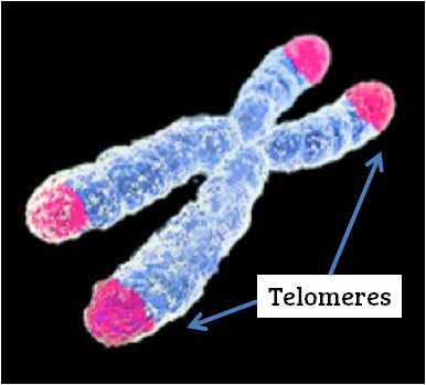
Telomeres are repetitive DNA sequences at the end of every chromosome.
They’re present to extend the life of our DNA, because every time a cell divides and its DNA is copied, a tiny piece of DNA is lost at the ends of chromosomes. So rather than losing a piece of important DNA, we lose a piece of the unimportant telomere.
ROS can damage DNA and cause the telomere to shorten, which can induce cellular senescence. Studies have shown that damaged, or shortened, telomeres can cause embryo arrest (Betts et al. 2008).
p66Shc
The p66Shc protein is a well-studied protein in cancer biology that can induce apoptosis/cellular senescence in response to damage by ROS.
It’s also been found in high levels in arrested bovine (cattle) embryos and these levels increase the longer the embryo is arrested (Betts et al. 2008). By removing p66Shc, this reduces embryo arrest and promotes blastocyst development (Betts et al. 2014).
Lab conditions
Embryos develop in the IVF lab before being transferred back to the uterus. The conditions that the lab provides are important to the embryo’s development and if they’re non-ideal, this might cause damage to the embryo and induce embryo arrest.
Oxygen tension
The air we breathe is about 21% oxygen but in the female reproductive tract the oxygen levels are between 2-8% (Morin 2017). This reduction in oxygen may be important in preventing ROS formation.
There is some controversy, but the general opinion is that embryos that are cultured in 5% oxygen tend to have higher success and are less likely to arrest (Morin 2017).
For economic reasons, some labs may still be culturing at atmospheric (21%) oxygen and this might increase embryo arrest.
Air quality
IVF labs need to have very clean air and are often fitted with heavy duty HEPA filters to purify the air.

Volatile organic compounds (VOCs) are molecules in the air that can be harmful to embryos. Some common VOCs are:
- Ethanol, isopropyl alcohol (for decontaminating surfaces)
- Propene, acetonitrile, styrene (from plastics)
- Formaldehyde, acetaldehyde (found in paints, gasoline, smog)
Having a clean lab and clean air is important for embryo health. For this reason strong smelling odors in deodorants or perfumes are not permitted. Plastic ware that is used for culturing is “degassed” before use by opening the packaging and letting the VOCs gas off.
Some VOCs are known carcinogens and can cause DNA damage.
Because embryos are so small and limited in how they can handle these toxins, VOCs can have big impacts on their development. VOCs have been linked to failure to produce blastocysts as well as reduced overall success (Mortimer et al. 2018).
Influence of sperm
Let’s look at some sperm-specific contributions to embryo arrest.
The centrosome
The centrosome is a structure inside cells that is needed to separate chromosomes during cell division. Chromosomes are separated by long fibers called spindle fibers that grab onto chromosomes and pull them apart. The spindle fibers originate in the centrosome. You can see this below:

Amargant et al. (2021) found that transferring sperm tails, which contain the centrosome, into activated egg cells allowed more embryos to develop to the morula stage compared to controls. In other words, eggs that received the centrosome from the sperm were less likely to arrest.
Burruel et al. (2014) took ROS-damaged sperm and injected it into rhesus monkey eggs, which all developed into embryos that arrested at the 8-cell stage. They found disorganized spindles in the cells, which they suggest might be affected by ROS-damaged centrosomes.
DNA fragmentation
Damage to the DNA in sperm can lead to sperm DNA fragmentation (SDF), as a result of environmental or lifestyle choices which may lead to increased ROS production.
Szabó et al. (2023) found that varicocele, impaired glucose tolerance, testicular tumors, smoking, pollution, and paternal age of over 50 were associated with the highest SDF. Some of these factors may be associated with oxidative stress due to increased ROS production, which has been associated with increases in SDF (Iommiello et al. 2015).
Sedó et al. (2017) found that there was a reduction in blastocyst formation rates in embryos from men with high levels of SDF, which was also associated with reduced pregnancy rates. Ni et al. (2014) also found a drop in blastocyst formation rates in the high SDF group, however this had no impact on pregnancy outcomes.
Adiga et al. (2021) performed a meta-analysis and combined the results of 23 studies on the impact of SDF and blastocyst formation. Overall, they concluded that there was no association with blastocyst formation and high/low SDF.
The biggest problem in this meta-analysis was how variable all the data in the 23 studies were, in particular the different types of SDF tests that were used (SCD, TUNEL, COMET) and the cutoffs for high/low SDF. This led the authors to state that the results “need to be interpreted with caution,” and better designed studies are needed with standardized threshold cutoffs.
Further adding to the variability is the impact of egg’s DNA repair system. Sperm can’t repair damage to its DNA, but the egg can.
Setti et al. (2021) found that there was no difference in blastocyst formation rates in women 40 or less with low/high SDF sperm used during IVF, but there was a difference when women were over 40. The authors suggest that this might be due to lower DNA repair activity in older eggs compared to younger eggs.
You can read the full summary of this study in my post Sperm DNA fragmentation and advanced female age.
How to correct embryo arrest
Unfortunately, embryo arrest is pretty common, and many patients will experience it during their IVF cycle. It will be more apparent with a higher number of eggs retrieved, but patients with fewer eggs may encounter it also.
While it may seem concerning if all embryos arrest when only a few are created, this can sometimes happen by chance. However, if multiple cycles show consistent embryo arrest, or if a high proportion of embryos arrest, this may indicate an underlying issue, and genetic testing could help identify possible causes.
There’s a lot of potential ways for embryos to arrest, as shown in this post:
- Errors with maternal effect genes
- Errors in the SCMC
- Errors in embryonic genome activation.
- Embryo aneuploidy
- Mitochondrial dysfunction
- Increased ROS levels due to environmental/lifestyle factors
- IVF lab conditions
- Sperm DNA fragmentation
Some factors may be potentially modifiable, such as certain lifestyle factors, while others may require different approaches like egg donation, sperm donation, or mitochondrial donation. Currently, there’s limited evidence for interventions that directly correct embryo arrest, aside from some early research on mitochondrial donation discussed earlier in this post.
Conclusions
Embryo arrest is when an embryo stops its development, and this usually occurs around day 3 which results in embryos failing to make it to blastocyst.
An important step takes place around day 3 when the embryo activates its DNA to produce the factors it needs to continue its development. Before this, the embryo relies on stored factors in the egg. Some embryos arrest because they have low levels of these factors, while others have problems in switching to the embryo’s DNA.
The subcortical maternal complex is a protein complex that’s involved in making this switch, and some cases of repeated embryo arrest are due to mutations in this complex.
Besides errors in switching to the embryo’s DNA, the embryo might have other problems that can induce embryo arrest, including aneuploidy, mitochondrial dysfunction or increased oxidative stress due to elevated ROS.
In some cases, non-ideal lab conditions can contribute to embryo arrest.
Sperm DNA fragmentation may also be a source for embryo arrest, although the evidence surrounding this is unclear and better studies need to be conducted.
Correcting embryo arrest may be possible through egg, sperm or mitochondrial donation. Other interventions may be possible, but more research is needed.
💡 Want to see real success stories with people who experienced a high degree of embryo arrest?
Check the IVF Success: Embryo arrest collection from the Uterine Wall of Fame, which features a growing collection of IVF success stories submitted by people who have been through it themselves.

About Embryoman
Embryoman (Sean Lauber) is a former embryologist and the founder of Remembryo, an IVF research and fertility education website. After working in an IVF lab in the US, he returned to Canada and now focuses on making fertility research more accessible. He holds a Master’s in Immunology and launched Remembryo in 2018 to help patients and professionals make sense of IVF research. Sean shares weekly study updates on Facebook, Instagram, and Reddit regularly. He also answers questions on Reddit or in his private Facebook group.