In a 2025 study, researchers tested a noninvasive MRI-like tool that reads an embryo’s metabolic health, accurately predicting which would stop developing and potentially helping select the healthiest for transfer.
Selecting the best embryo for transfer is one of the most important steps during IVF. There are a number of tools that aim to rank embryos for transfer, including morphological grading, time-lapse imaging, and PGT-A, but they tell us little about an embryo’s metabolism, or the chemical processes that help the embryo grow and develop.
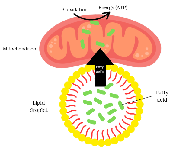
A key part of this metabolism happens in the mitochondria, often called the “powerhouses” of the cell. Mitochondria turn nutrients into ATP, the cell’s main energy currency. While they can make ATP from sugars, they can also make it from fats through a process called β-oxidation.
That’s where lipid droplets come in. These are tiny storage bubbles inside cells that hold fatty acids, which are a dense and energy rich fuel. When needed, lipid droplets release these fatty acids, which are transported into mitochondria and broken down to produce ATP. This stored energy is especially important in early embryos, which can’t yet rely on the mother’s blood supply for nutrients.
Measuring this kind of internal energy reserve could be just as important as assessing embryo structure or genetics, but it has been difficult to do without harming the embryo. Most “noninvasive” approaches only analyze molecules released by the embryo into the culture medium, giving an indirect and often incomplete picture of the embryo’s metabolic health.
In a new study by Sivelli et al. 2025, researchers test a new technique called micro magnetic resonance spectroscopy (micro MRS) that can detect fatty acids stored in lipid droplets. These droplets can be a marker of the embryo’s energy balance: too few may limit growth, while too many can signal metabolic stress. The micro MRS measures the chemical properties of the lipid droplets inside the embryo, creating a kind of molecular signature that can give information about the embryo’s energy balance and overall health.
The technique works on the same basic principles as an MRI, a hospital imaging tool that uses strong magnets and radio waves to create detailed pictures of the inside of the body. Instead of producing an image, micro MRS measures the chemical fingerprint of the embryo, based on the signals it detects from certain molecules inside cells.
🔗 Original studies are referenced in this post or within the linked Remembryo posts.
💡 Reminder: Terms underlined with a dotted black line are linked to glossary entries. Clicking these does not count toward your paywall limit.
Micro MRS is highly accurate in predicting embryo arrest
Researchers tested whether micro MRS could give useful insights into embryo health by applying it to more than 150 bovine embryos and oocytes. Embryos at the 8 cell stage and oocytes were placed in the micro MRS device for about an hour, allowing the instrument to measure the chemical signature of their lipid droplets.
You can see a video of how micro MRS is done in the lab below (Sivelli et al. 2025, CC by 4.0):
In 8 cell embryos, they found 14 markers that differed between those that went on to develop to the expanded blastocyst stage and those that arrested before this point. A machine learning model correctly ranked embryos by their likelihood of developing about 94% of the time (AUC = 0.94), meaning it was highly accurate in predicting which embryos would later arrest.
In oocytes, several markers differed between immature and mature eggs, but the model was less accurate in this case, distinguishing between the two about 69% of the time (AUC = 0.69).
These findings suggest that micro MRS can detect chemical differences in embryos and eggs that relate to their developmental potential. If confirmed in human studies, this approach could become a useful, noninvasive tool for ranking embryos or eggs for IVF.
But is the technique safe?
Safety was assessed by exposing mouse embryos to the static magnetic field used for micro MRS, transferring them to surrogate mothers, and tracking health and reproduction over three generations. There were no differences between exposed and control embryos in implantation rate, live birth rate, offspring health or fertility, even in later generations, suggesting the process is safe in this animal model.
Conclusions
This study introduced micro MRS, a noninvasive tool that measures fatty acids stored in lipid droplets within embryos or egg cells to provide information about their metabolic health. In 8 cell embryos, the technique predicted with very high accuracy which ones would later arrest, and in oocytes it showed moderate accuracy in distinguishing mature from immature eggs. Safety tests in mouse models found no negative effects on embryo development, offspring health, or reproduction.
Each micro MRS measurement takes about an hour and produces a metabolic profile that could help embryologists assess quality without taking a biopsy. Human studies will be needed to confirm whether these measurements can predict implantation or live birth, but the ability to detect embryo health through metabolic signatures is promising. In the future, the technique might not only identify embryos likely to arrest but also help rank developing embryos by their potential for a successful pregnancy.
The authors note that other noninvasive tests, like ni-PGT that analyzes the culture media embryos have been in, aren’t very accurate because they only measure what the embryo releases or absorbs, not what’s inside it. This means important internal markers can’t be assessed, and the tiny size of the embryo compared to the large volume of culture media makes useful signals hard to detect.
However, it’s not clear if this technique could be a good replacement for PGT-A, since micro MRS is not evaluating the embryo’s chromosomes. Still, it would be interesting to see if there’s any links between what micro MRS finds and PGT-A.
Limitations of this research include the fact that it was conducted in cow and mouse embryos rather than humans, and the cattle embryos had been cryopreserved at an early stage, a step known to reduce survival in that species. The number of embryos that developed was small, and the measurements were performed without gas control, although temperature was maintained.
For more reading on the subject, check out these posts:
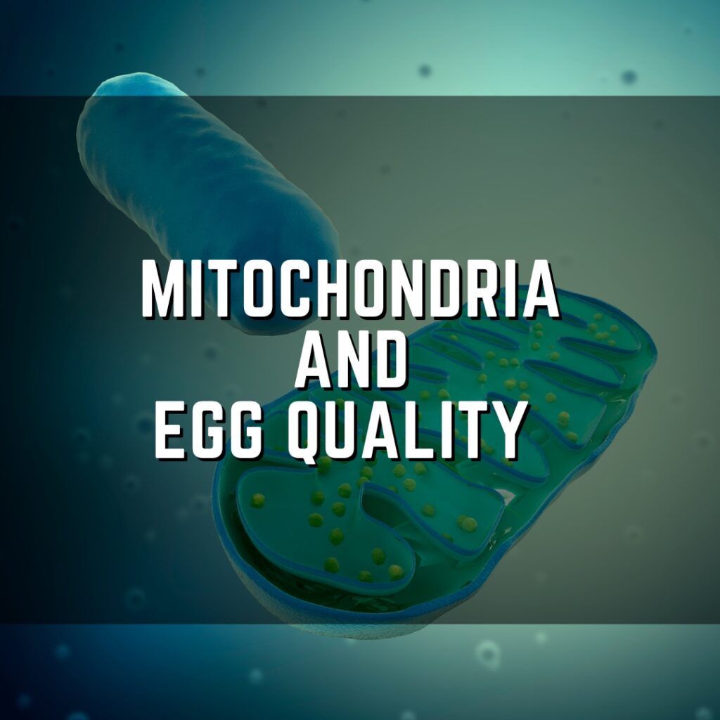
The egg cell contains a high number of mitochondria, which are tiny cellular organs that produce energy that the egg needs to power fertilization and embryo development. Research has shown that these mitochondria can become dysfunctional with age, leading to reduced energy availability for the egg that could result in poor egg quality. Different treatments that target mitochondria, like antioxidant supplements or mitochondrial donation, may improve egg quality, although good quality research is lacking that shows these treatments are truly effective. Read more.
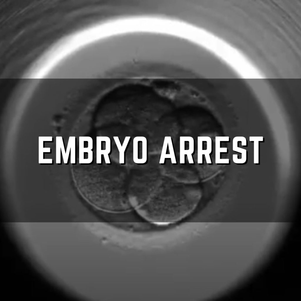
Embryo arrest is when an embryo stops developing, usually before reaching the blastocyst stage. This post explains possible causes, including embryonic genome activation, maternal effect genes, the subcortical maternal complex, aneuploidy, mitochondrial dysfunction, oxidative stress, lab conditions, and sperm DNA fragmentation. Read more.
Reference

About Embryoman
Embryoman (Sean Lauber) is a former embryologist and the founder of Remembryo, an IVF research and fertility education website. After working in an IVF lab in the US, he returned to Canada and now focuses on making fertility research more accessible. He holds a Master’s in Immunology and launched Remembryo in 2018 to help patients and professionals make sense of IVF research. Sean shares weekly study updates on Facebook, Instagram, and Reddit regularly. He also answers questions on Reddit or in his private Facebook group.
Related posts:
 Embryo Arrest
 Pilot study shows improved IVF outcomes after spindle transfer in women with poor egg quality
 Aneuploidy is a key factor in embryo arrest
 Scientists identify genes linked to unexplained infertility and repeated IVF failure
 Complete Guide to Embryo Grading and Success Rates
 Day 3 or Day 5 embryo transfer?
 Predicting how many day 3 embryos make it to blastocyst
 Blastocyst and pregnancy rates of day 3 embryos based on cell number, fragmentation