
Researchers in a 2022 study found that the combination of endometriosis and adenomyosis results in lower cumulative live birth rates after IVF, particularly in those with T2 high-signal intensity myometrial spots as evaluated by MRI.
Adenomyosis is characterized by the presence of endometrial-like tissue in the myometrium, the muscular part of the uterine wall that lies between the endometrium and the perimetrium.

Adenomyosis can be diagnosed by transvaginal sonography (TVS) and magnetic resonance imaging (MRI). You can see an example of a pelvic MRI below showing adenomyosis in an enlarged myometrium.

Adenomyosis is found in a large proportion of infertile women (24.4%, Puente et al. al 2016) and can be associated with endometriosis in about 20-80% of patients (Vannuccini et al. 2019). It’s not clear how adenomyosis affects fertility, however it may have to do with changes in how the endometrium functions, inflammatory reactions, or how the uterus contracts to transport sperm (Sharma et al. 2018).
🔗 Original studies are referenced in this post or within the linked Remembryo posts.
💡 Reminder: Terms underlined with a dotted black line have glossary definitions. Hover or tap to see a short definition, or select “Read full definition” in the pop-up to view the complete glossary entry. Viewing glossary entries does not count toward your paywall limit.
In this study, Bourdon et al. (2022) followed 202 women (median age of 33.0) with endometriosis to determine the frequency and impact of adenomyosis on live birth rates. Most of these cases were severe, with 90.1% having deeply infiltrating endometriosis, and 72% of these women having ovarian endometriomas (OMA).
Of these 202 women, 145 (71.8%) had adenomyosis that was diagnosed using pelvic MRI, and 57 did not.
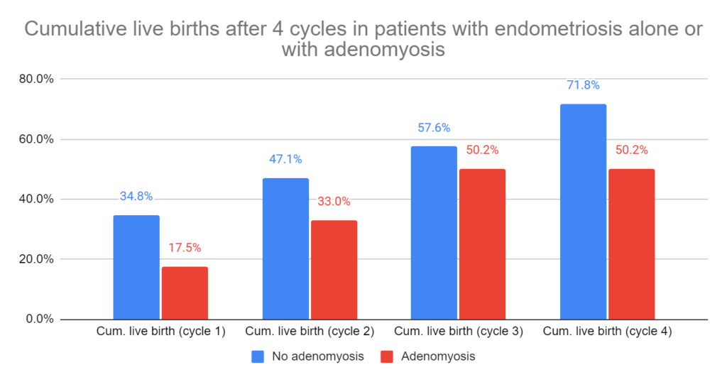
This study examined cumulative live birth rates in these 202 women who a total of 346 IVF cycles, with some women doing up to 4 cycles. The women with adenomyosis had a lower cumulative live birth rate compared to the women without it (50.2% vs 71.8% over 4 IVF cycles).
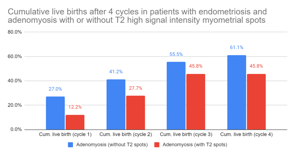
Of the 145 women with adenomyosis, 44.1% of them had “T2 high-signal intensity” myometrial spots. This refers to how the spots appear under MRI and may represent microcysts of adenomyosis. These women had 5 of these spots on average. Cumulative live birth rates for women with these T2 high-signal intensity myometrial spots were lower compared to women without them (45.8% vs 61.1% after 4 cycles).
They also performed a multivariate analysis and found that the presence of adenomyosis, and the presence of the T2 high-signal intensity myometrial spots, both independently resulted in lower live birth rates (as did age and AMH levels). This tells us that age, AMH levels, the presence of adenomyosis, and the presence of T2 high-signal intensity myometrial spots, all independently correlated with live birth rates.
This study is supported by another study by Sharma et al. (2019) that also found a decrease in live births with endometriosis and adenomyosis. The difference is that this study used MRI to evaluate the impact of these T2 high-signal intensity myometrial spots, and found that having more of them leads to worsened outcomes. Counting these lesions should be possible when a pelvic MRI is performed and, as this study demonstrates, the presence of these spots leads to a worse prognosis.
Treatment for adenomyosis is mostly lacking, and may involve surgery (MRI-guided thermal ablation, myomectomy, hysterectomy) or therapeutics (oral contraceptive pills, levonorgestrel intrauterine device, danazol, aromatase inhibitors), although the evidence surrounding their effectiveness and potential risks is not clear (Gunther et al. 2022).
Reference

About Embryoman
Embryoman (Sean Lauber) is a former embryologist and the founder of Remembryo, an IVF research and fertility education website. After working in an IVF lab in the US, he returned to Canada and now focuses on making fertility research more accessible. He holds a Master’s in Immunology and launched Remembryo in 2018 to help patients and professionals make sense of IVF research. Sean shares weekly study updates on Facebook, Instagram, and Reddit regularly. He also answers questions on Reddit or in his private Facebook group.